 In this shot (L to R): Centrifuge, Microtubule tray, Pipettes on holder, box of gloves, PCR, Heater pad, Water Bath
Friday, June 19, 2009
Uncluttered Bench - Long view
 In this shot (L to R): Centrifuge, Microtubule tray, Pipettes on holder, box of gloves, PCR, Heater pad, Water Bath
PCR - Basic
PCR - Plus
 In the shot: PCR machine, pipettes, enterotube, petri dishes
In the lab but not used with PCR: The other items are all reasonable candidates to add to the same bench in this type of lab.
Uses: DNA analysis when you've only got a little blood... You can make lots of copies of a specific piece of DNA.
Labs use Polymerase chain reaction (PCR) machines to detect and amplify a specific DNA sequence against the background of a complex genome. This procedure is very powerful because it can be used on fragments of DNE that are initially present in infinitesimally small quantities.
Tissue Staining - Basic
Tissue Staining - The Rinse
Dissection Scope - Basic
 In the shot: Dissection scope, scalpel, petri dish with bacterial culture. Dissection work is often done near a sink.
 Options: Add a sink, dish soap and latex gloves nearby. Bits of bone, quarter size piece of tissue being dissected, plant material, larger items to be magnified.
Additional: camel hair brush, morgue (small bottle with water and detergent), culture vials (square bottles), deionized water, label tape, markers, white cards, scissors, anaesthetizing vial (round bottles) add if you are using live insects, etc.
Compound Microscope Tray
Compound Microscope - Basic
Thermal Heater - Basic
CENTREFUGE - UNBALANCED
Electrophoresis - Plus
 In this shot: Electrophoresis analytic tool, slides, notebook, gel
Add: Buffer, test tubes, brand name stains like SimplyBlue™ SafeStain and SYPRO® Ruby are used for detection of proteins in gels.
Electrophoresis is an analytical tool for separating proteins and nucleic acids. An electric current is passed through a medium containing the target mixture, and each type of molecule travels through the medium at a different rate, depending on its electrical charge and/or size. Agarose and acrylamide gels are the most common media for electrophoresis of nucleic acids and proteins, respectively.
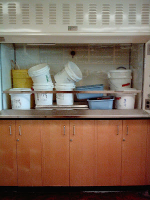
FUMEHOOD - AS STORAGE
Petri Dish - E. Coli
Labware Labelling

In this shot: Flask with hand-written label
Hand-labelling is common in labs. Any beaker or flask can be labelled with red, blue, orange, pink, yellow or white tape.
Common labels clear liquids: 0.1 gr/1000 ml TETRA lolium (toxic)
H2So4, CH4SO4, Acidified H2O2, 0.0125 M HCL
Common labels on Brown (liteSafe) bottles:
Iodine, Carolina BLU DNA Stain, Phosphoric Acid (85%)
Laboratory Chemicals
Subscribe to:
Comments (Atom)


